

ECHO
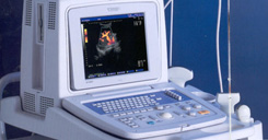
What is an Echocardiogram: An echocardiogram is a test in which ultrasound is used to examine the heart. The equipment is far superior to that used by fishermen. In addition to providing single-dimension images, known as M-mode echo that allows accurate measurement of the heart chambers, the echocardiogram also offers far more sophisticated and advanced imaging. This is known as two- dimensional (2-D) Echo and is capable of displaying a cross-sectional "slice" of the beating heart, including the chambers, valves and the major blood vessels that exit from the left and right ventricle
An echocardiogram can be obtained in a physician's office or in the hospital. For a resting echocardiogram (in contrast to a stress echo or TEE, discussed elsewhere) no special preparation is necessary. Clothing from the upper body is removed and covered by a gown or sheet to keep you comfortable and maintain the privacy of females. The patient then lies on an examination table or a hospital bed
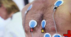
Sticky patches or electrodes are attached to the chest and shoulders and connected to electrodes or wires. These help to record the electrocardiogram (EKG or ECG) during the echocardiography test. The EKG helps in the timing of various cardiac events (filling and emptying of chambers). A colorless gel is then applied to the chest and the echo transducer is placed on top of it. The echo technologist then makes recordings from different parts of the chest to obtain several views of the heart. You may be asked to move form your back and to the side. Instructions may also be given for you to breathe slowly or to hold your breath. This helps in obtaining higher quality pictures. The images are constantly viewed on the monitor. It is also recorded on photographic paper and on videotape. The tape offers a permanent record of the examination and is reviewed by the physician prior to completion of the final report